
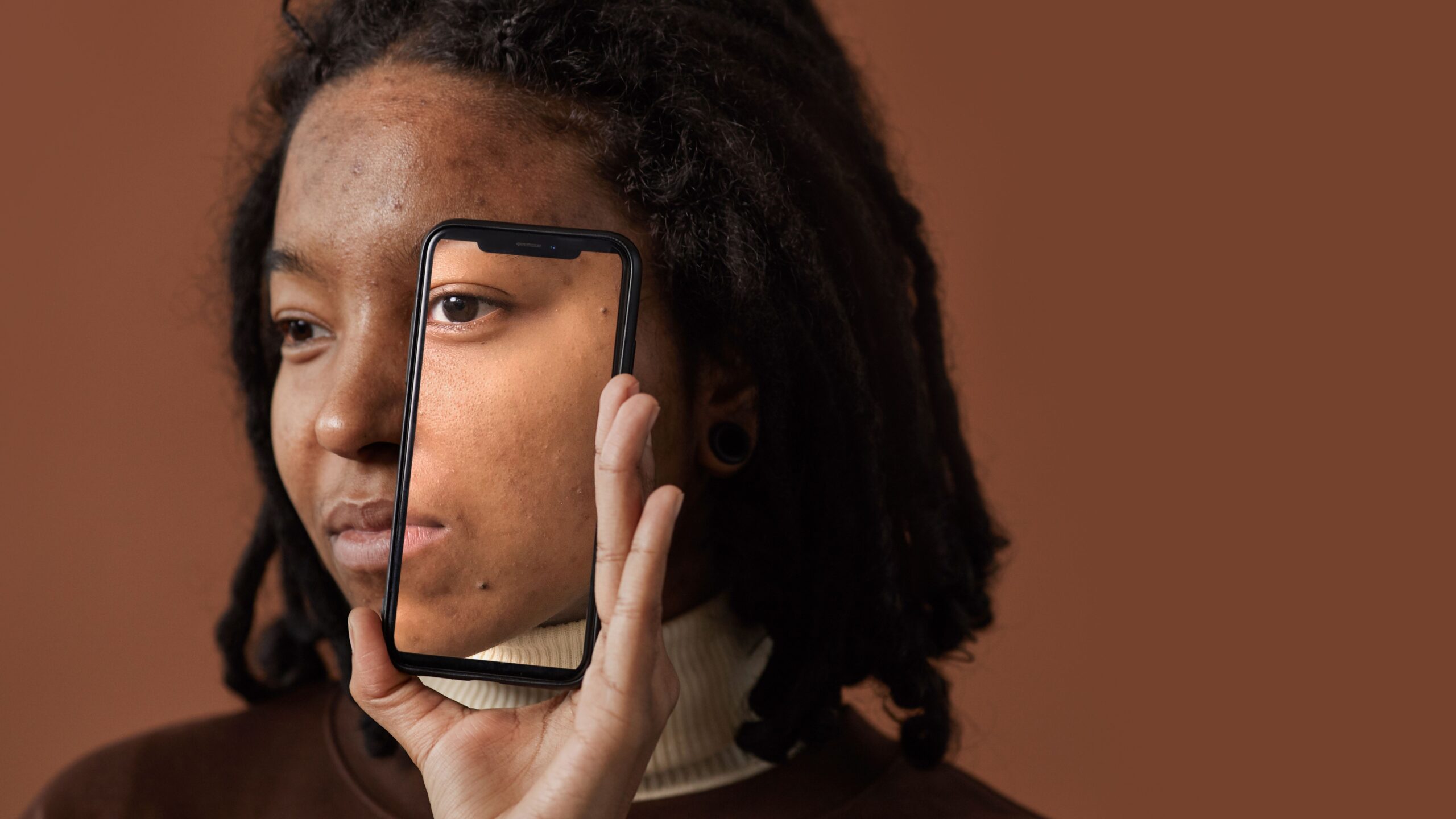
Two distinct tiers of skin analysis technology exist today, and each one captures a different depth of information about your skin. An AI phone app reads what a camera sensor can see under regular light. A professional-grade system goes further still, layering multiple light sources together to reveal what’s happening beneath the surface.
Two Tiers of Skin Analysis Technology
| Tier | Hardware | Light sources | Typical setting |
|---|---|---|---|
| Consumer AI apps | Phone camera | Visible light only | Home |
| Clinic-grade systems | Multi-camera imaging rig | White, UV, cross-polarized, RBX | Dermatology offices |
Each tier trades off convenience against depth. AI app takes seconds and costs nothing, but works with a single visible-light image. A clinic system takes longer, usually requires an appointment, and captures several image types in one sitting.
Decoding the Light Spectrum Behind a Scan
Different light sources expose different layers of skin, and understanding this is the key to reading any skin analysis report:
- Standard/white light captures surface texture, visible spots, and general tone, roughly matching what you’d see in a well-lit mirror
- UV light reveals sun damage that hasn’t surfaced yet, along with porphyrins, a fluorescent marker linked to acne-causing bacteria
- Cross-polarized light filters out surface glare to show redness and vascular patterns sitting beneath the skin’s top layer
- RBX imaging separates red and brown pigment signals from each other, giving a clearer picture of whether a discoloration is vascular or melanin-based
None of these light sources work in isolation on a clinic-grade device. They’re layered together into one composite report, which is part of why a single dermatology visit can surface findings a bathroom mirror never would.
Inside Clinic-Grade Systems Like VISIA
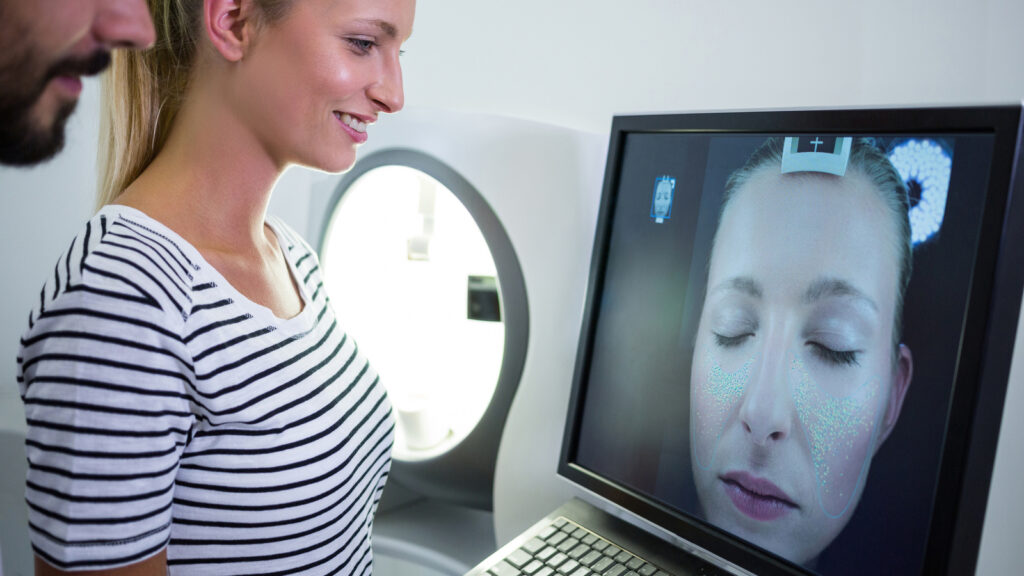
VISIA, made by Canfield Scientific, is the imaging system most commonly referenced as the industry benchmark in dermatology offices and med spas. The system captures multiple facial images using several cameras and light sources in a single session, then scores results across categories including spots, wrinkles, texture, pores, UV spots, brown spots, red areas, and porphyrins. Results are typically compared against a database of prior scans grouped by age, giving a percentile-style readout rather than a raw, context-free score.
A typical session takes only a few minutes. You position your face against a chinrest, the system captures images from multiple angles in quick succession, and the software generates a report almost immediately afterward. Because each scan is stored, providers can pull up a previous session months or years later and compare it directly against a new one, which is part of why these systems are used to track visible progress from a skincare routine or in-office treatment over time, not just to produce a one-time snapshot.
Accuracy Across the Tiers
Independent validation exists for VISIA specifically, and the numbers are more concrete than most think:
- A 2022 study (published in GMS Interdisciplinary Plastic and Reconstructive Surgery) tested VISIA’s precision across repeated scans and found differences under 2% for texture, UV spots, brown spots, and porphyrins, with pores and red areas landing between 2% and 4%.
- A 2023 follow-up study found VISIA’s calculated “skin age” correlated strongly with participants’ real age, with age estimates typically landing within 3 to 5.5 years of a person’s actual age.
Consumer AI apps tell a different story. Board-certified dermatologists interviewed by LovelySkin have cautioned that phone-based face analyzers can produce inconsistent results and shouldn’t replace an in-person exam, pointing to broader concerns about apps operating without FDA oversight. Clinical AI comparisons from Haut.AI note that research-grade tools measure specific parameters like redness or pore density with real precision, while consumer-facing apps often trade some of that rigor for speed. In practice, many consumer tools are benchmarked against VISIA-style measurements in the first place, which means the gap between tiers usually comes down to how much independent, published testing a specific product has gone through, rather than a fundamentally different technology underneath.
Consumer App or Professional System: What Changes
- Depth of imaging — a phone photo captures one light condition; a clinic session captures several
- Interpretation — an app returns a generated report; a professional session pairs results with a person trained to interpret them and recommend next steps
- Validation — clinic systems like VISIA have published precision data behind them; app-level accuracy claims vary and are harder to verify independently
- Cost and access — apps are free or near-free and instant; professional sessions require an appointment and typically cost more
Reading Your Results Without Overreacting
A skin analysis report, from any tier, works best as a starting point rather than a diagnosis. Use it to spot patterns worth paying attention to: consistently high UV-spot scores might nudge you toward better sun protection habits, and elevated porphyrin readings might explain recurring breakouts in a specific area. If your report flags noticeable redness or vascular patterns under cross-polarized light, that’s useful context for choosing barrier-supporting ingredients rather than reaching for something stronger. The goal is matching a category of concern to a category of solution, not treating any single score as a verdict on your skin’s overall health.
Flags that involve asymmetric moles, rapidly changing spots, or anything that looks unusual beyond typical sun damage or texture scoring deserve a dermatologist visit rather than a shopping list, since no analysis device, consumer or clinical, is built to diagnose skin cancer.
🧪Lab Verdict
Skin analysis devices span a wide range, from a free phone scan to a multi-camera clinic system with published precision data behind it. Clinic-grade tools like VISIA carry the strongest independent validation currently available, particularly for wrinkle scoring and age estimation. Consumer apps offer a convenient starting point, but the accuracy varies by product, and dermatologists still recommend an in-person visit for anything beyond general skincare curiosity. Treat any tier’s results as a guide for adjusting your routine, not a substitute for a professional opinion when something looks off.
FAQ
Are skin analysis apps as accurate as clinic scanners?
Not consistently. Clinic-grade systems like VISIA have published precision studies behind them, while consumer app accuracy varies by product and hasn’t undergone the same level of independent testing.
How often should you get a professional skin analysis?
There’s no specific rules, but many dermatology offices and med spas suggest revisiting every few months to a year to track changes over time, especially if you’re adjusting your skincare routine.
Can a skin analysis device detect skin cancer?
No. These devices are built to score cosmetic concerns like texture, spots, and pigmentation, not to diagnose disease. Any unusual mole or lesion should be evaluated by a dermatologist directly.
Leave a Reply